
 Français
 Español
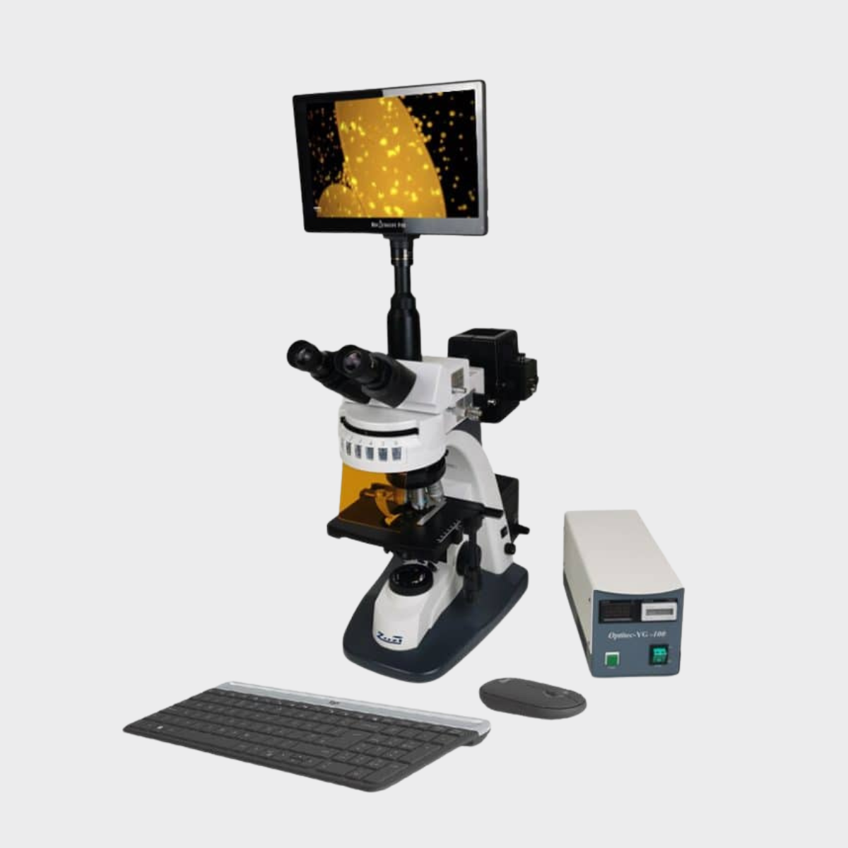


Products

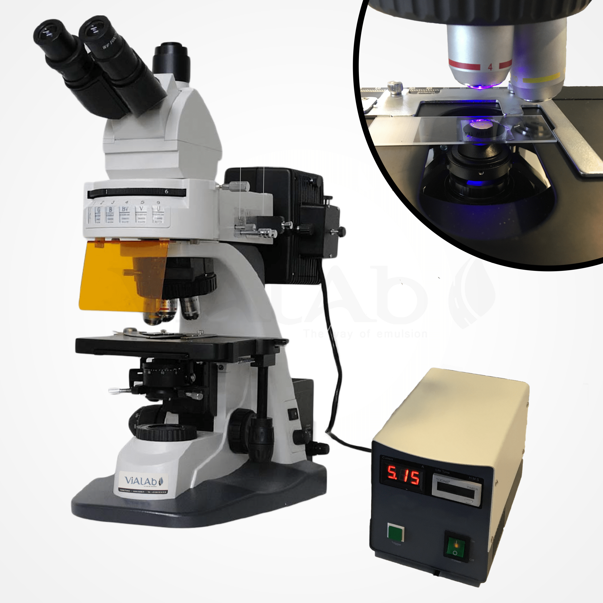
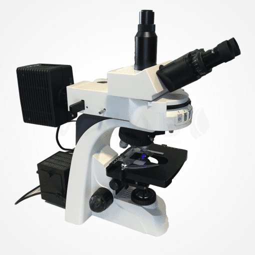
Epi-fluorescence Microscope | Touchscreen Tablet 11.8" | 16MP Integrated Camera
Technical features
- Tip: Triocular, 0-40-degree inclination with adjustment of inter-pupil distance: 50-75 mm and dioptric correction
- Eyepiece WF 10x/20
- Sextuple barrel with apochromatic fluorescence plane lenses 4x (0.15); 10x (0.35); 40x (0.75); 100x (0.90)
- Double platinum 180 x 160 mm mechanical displacement 80 x 50 mm
- Transmission: Halogen lamp 12V, 50W
- Epi-fluorescence: High-pressure mercury lamp 20V, 100W
- Illumination type Köehler with field diaphragm and aperture
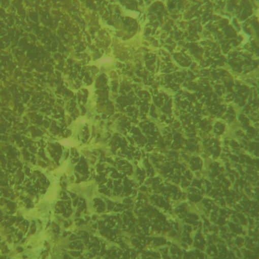
- 5 Excitement Filters (EX) / Emission (EM):
- Green EX 510-560 – EM 590 nm,
- Blue EX 450-490 – EM 520 nm,
- Blue/Violet EX 400-440 – EM 470 nm,
- Violet EX 380-420 – EM 450 nm,
- Ultra-Violet EX 330-380 – EM 420 nm
- Touch tablet with built-in camera and image acquisition software:
- Touch screen 11.8″
- Resolution 4032×3024 pixels
- 16-megapixel camera
- Pre-installed image acquisition software (photos and videos) and analysis (measures of lengths, surfaces,…)
- Inlay of scales in photos
- Windows 10 operating system
- Wifi
- Intel Atom 1.44 GHz processor
- 4GB Ram
- 60GB storage space
More information
- Mice and wireless keyboard via USB 2.0 port provided
- Food: 3×230 VAC, 50 Hz
- Epifluorescence feeding
- General power for the microscope and its halogen lamp
- Tablet power with DC 12V transformer
- Spare part:
Mercury high-pressure lamp 20 V 100 W. (Limited lifespan 200h)
F044.136.2030
Accessories & Options

Microscope Glass Slide | 76x26 mm | Pack of 50
- Thickness: 1 / 1.2mm
- Dimensions: 76 × 26mm
- White band identification
F039.136.2020

Microscope Glass cover slide | 24x40 mm | Pack of 500
- Thickness: 0.13 / 0.17 mm
- Dimensions: 24 × 40 mm
- Borosilicate glass
F039.136.2021